


Visual Field Test
Visual field tests assess the potential presence of blind spots (scotomas), which could indicate eye diseases. A blind spot in the field of vision can be linked to a variety of specific eye diseases, depending on the size and shape of the scotoma.
Many eye and brain disorders can cause peripheral vision loss and visual field abnormalities.

For example, optic nerve damage caused by glaucoma creates a very specific visual field defect. Other eye problems associated with blind spots and other visual field defects include optic nerve damage (optic neuropathy) from disease or damage to the light-sensitive inner lining of the eye (retina).
Brain abnormalities such as those caused by strokes or tumors can affect the visual field. In fact, the location of the stroke or tumor in the brain can frequently be determined by the size, shape and site of the visual field defect.
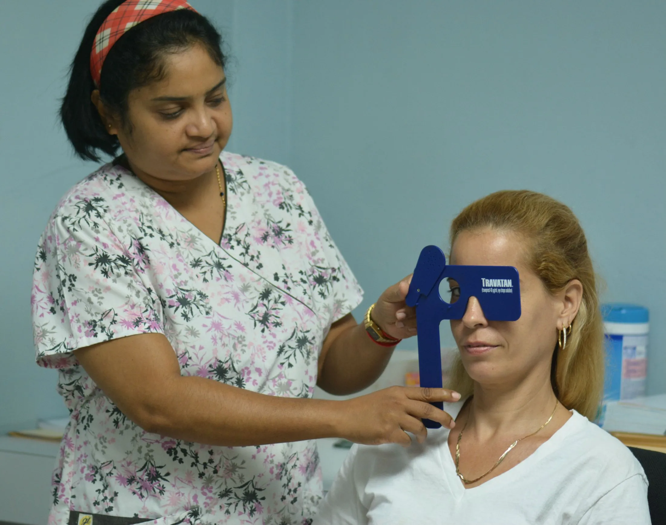
 Confrontation visual field testing typically is used as a screening visual field test. One eye is covered, while the other eye fixates on a target object, such as the doctor’s open eye, while the doctor stands or sits directly in front of you. You then are asked to describe what you see on the far edges or periphery of your field of view.
- Automated Perimetry. Various forms of automated perimetry tests measure your responses to the presence of objects in different areas of your field of view.
While your head is held still, usually with a chin rest inside a large bowl-like instrument, you stare at a source of light straight ahead. Random lights of different intensities are flashed in your peripheral field of vision.
You then press a button or use other means to indicate your response when you perceive the computer-generated light suddenly appearing in your field of view.
If you can’t see objects in an appropriate portion of your field of view, then you may have a blind spot indicating vision loss. - Frequency Doubling Perimetry. Frequency doubling is based on an optical illusion produced with vertical bars of contrasting colors (usually black and white) appearing on a screen. These bars appear to double in number when they alternately flicker at higher frequencies, a phenomenon thought to be due to the unique response of specific light-sensitive cells (photoreceptors) in the retina.
Inability to see vertical bars at certain frequencies could indicate optic nerve or other types of eye damage with accompanying loss of vision in certain areas of the visual field. - Electroretinography. This test measures electrical activity generated by the photoreceptor cells in the retina when the eye is stimulated by a special strobe light or a reversing checkerboard pattern of light. The measurement is captured by an electrode placed on the front surface of the eye (cornea), and a graphic record called an electroretinogram (ERG) is produced.